ESVD Research Grants
The ESVD research grants
These grants will be awarded by the ESVD for basic or clinical research in veterinary dermatology. Applicants will be expected to propose a project of scientific merit that is applicable to veterinary dermatology. The projects are expected to be of one to two year’s duration. Preference will be given to novel proposals including the development of pilot studies, but applications of ongoing research work will be also considered.
- Research proposals together with a separate curriculum vitae of each of the principal investigators should be submitted by April 1st 2024 midnight CET to publications-grants@esvd.org
- Additional information about the application can be found here
Please send your complete application to publications-grants@esvd.org
Types of research grants
two MAJOR GRANTs - €15,000
for an outstanding advanced research project
two MINOR GRANTs- €8,000
for a starting researcher or private practicioner
Selection Process
Grants are evaluated by the grant awarding committee on scientific merit, feasibility, and usefulness. The successful applicant(s) will be informed as soon as a decision has been reached and the successful project applications are announced at the following AGM of the Society.
Conditions
Annual progress reports are required and should be submitted to the current secretary at least 30 days before the Society’s AGM. Successful applicants are encouraged to submit their results for publication in the Journal of Veterinary Dermatology and to present their findings to ESVD members at the annual congress. Payment of the grant will be made into a special account and a budget report will be required by the ESVD Treasurer at the end of the study. Any funds not spent must be returned to the ESVD The funds cannot be used for travel and accommodation at the meeting but purely for research. However, the principal awardee is entitled to one free registration at any annual ESVD congress (this does not include the WCVD) at which he or she will present data generated as a result of the grant.
Information about grants awarded
The ESVD Research grants 2023 were awarded the following researchers:
Major grant (€15.000): Dr Carolina Frizzo Ramos "Ameliorating Canine Atopic Dermatitis by targeted micronutrition"
Training grant (€5.000): Dr Nikoleta Makri "Correlating DNA sequencing, culture and cytology to evaluate the microbiome in healthy rabbit ears
Minor grant (€5.000): Dr Matt McHale "In vitro antimicrobial efficacy and in vivo residual activity of a 3% chlorhexidine shampoo and foam in healthy dog"
Major grant 2023
Grant receivers: Carolina Frizzo Ramos, Franziska Roth-Walter, Lucia Panáková
Presentation of the study: Dysbalanced iron homeostasis is linked to inflammation, with the first description of reduced dietary
uptake of iron (‘mucosal block’) upon inflammation being described in dogs. As a skewed iron
homeostasis is associated with a greater morbidity and mortality in a number of chronic
inflammatory diseases in humans, here we aimed to circumvent the mucosal block existing in dog
patients suffering from canine atopic dermatitis (CAD) by using fortified whey. Dietary uptake of
micronutrient-enriched whey occurs via the lymph system and is not affected by the mucosal block
allowing targeted micronutrition.
Our preliminary assessment of iron parameter in healthy and dogs suffering from canine atopic
dermatitis that – similarly as in humans- iron homeostasis is affected in these dogs and is linked to
inflammation. Moreover, in one case study supplementing a French bull dog suffering from CAD
with micronutrient-enriched whey led to a sustained disappearance of wounds and itching and the
discontinuation of any medication since over 18 months
In a next step and with support of the ESVD, we aim here to assess whether 4 months of dietary
intake of micronutrient-rich whey can improve the immune status and reduce the inflammatory
symptom burden in CAD-patients in a randomized, double-blind, placebo-controlled manner.
Minor grant 2023:
Grant receiver Matt McHale
Presentation of the study:
This study is to assess four set contact time points for shampoo in healthy animals for in vitro antimicrobial activity using hair clippings, with one control time point (0 minutes). Residual activity would then be assessed after one application of the shampoo. Residual activity would also be assessed after one application of the mousse.
Once we have established the optimum shampoo contact time and foam interval for efficacy, we will then assess the in vivo efficacy with atopic dogs, with microbial overgrowth.
_______________________________________________________________________________________________________________
The ESVD Research grants 2022 were awarded the following researchers:
Major grant (€15.000): Dr A Lurenco
Training grant (€5.000): Dr N Apostolopoulos
Minor grant (€5.000): Dr V Schmidt
_______________________________________________________________________________________________________________
Major grant 2021
"Use of bacteriophages in the treatment of suppurative canine otitis externa caused by multiresistant Pseudomonas aeruginosa"
Grant receivers: Jacques Fontaine, Caroline Leonard, David Thiry
Presentation of the study:
Selection and storage of Pseudomonas strains
Training grant 2021
“Histopathological examination of non-blanching erythematous dermatitis in the dog, a retrospective study”

Grant receivers: Stephanie Winter, Brett Wildermuth, Dr. Kerstin Wildermuth, Dr. Ralf Müller, Dr. Sonya Bettenay, Dr. Nadine Meertens
Presentation of the study: Diascopy is a useful tool in veterinary dermatology to evaluate erythematous dermatitis, which aids in forming a list of differential diagnoses. Erythema which blanches on diascopy is termed positive and indicates vasodilatation and non-blanching erythematous lesions (diascopy is negative) are due to haemorrhage into the skin. Diseases of the dog in the literature whereby diascopy is reported to be negative include epitheliotropic T-cell lymphoma, vasculitis, erythema multiforme, cutaneous sterile pyogranuloma, Wells'-like syndrome, neutrophilic dermatitis and panniculitis (Sweet’s syndrome) and drug induced dermatitis. Not all of these diseases are traditionally thought of as diseases with dermal haemorrhage. The aims of this retrospective study are to report which diseases are negative upon diascopy and to investigate for the presence or absence of haemorrhage and vascular changes upon histopathological examination.
Training grant 2021
"Detection of canine allergen specific IgE: Comparing the molecular-based macro array (ALEC chip) to conventional ELISA testing on allergen extracts (Allercept Heska) and their correlations to clinical symptoms"

Grant receivers: Nina Poláková, Lucia Panáková, Isabella Pali-Schöll
Presentation of the study:
Background: Because of known limitations in both, serum and intradermal allergy tests, none can be considered a gold standard in canine allergology. This is partially due to the lack of standardisation of allergen extracts. However, testing for allergen specific IgE using an ELISA based on Fc-ε receptor α- chain (further described as ELISA) has been shown to have a good intra- and interlaboratory repeatability.
Objectives/Hypothesis: The goal of the current pilot study is to investigate the utility of the molecular allergen diagnostic method ALEX macro array chip for allergy testing (detection of allergen-specific IgE antibodies) in canine atopic patients and to compare the results with ELISA. Client owned dogs with clinical diagnosis of canine atopic dermatitis will be included in the study. We hypothesise to see correlation between macro array and ELISA and clinical presentation of patients. The long-term goal is to establish a reliable and sensitive method for molecular allergy- testing in canine patients with a good correlation to clinical symptoms, while only small volume of serum for testing of a broad range of allergen-molecules and extracts is needed.
Practitioner grant 2021
"Retrospective study on poor coat condition and alopecia in Pomeranian dogs and an overview of clinical, electron microscopic and histopathological findings in healthy and affected Pomeranian dogs"
Grant receivers: Dr. Annette van der Lee, Kelly van Amersfort, Dr. Jaco van der Lugt
Presentation of the study: Acquired coat abnormalities are frequently seen in Pomeranian dogs, leading to great dissatisfaction of owners. Two distinct coat types exist within this breed; the soft shiny coat and the wooly coat. When alopecia occurs in the furthermore healthy Pomeranian dog, the diagnosis of Alopecia X is often made. Our hypothesis is that Alopecia X is not one single disease entity, but a progressive form of poor coat quality seen in the wooly coat type Pomeranian.
The aim of this study is to find risk factors for developing Alopecia X in the Pomeranian. By means of a retrospective epidemiologic study, the prevalence of Pomeranians with Alopecia X within the Netherlands will be estimated. Possible risk factors as coat type, coat color and gender will be evaluated to confirm our hypothesis. By means of a prospective cohort study collecting skin biopsies from affected alopecic (group 1) and non-affected wooly coat (group 2) and shiny coat (group 3) Pomeranians, histopathological findings will be compared. In addition, similarities of the wooly coat type with the human disorder pili trianguli et canaliculi will be explored. For this matter, scanning electron microscopy will be used to compare the structure of hairs of the three groups.
Major grant 2020
“Molecular characterization of the cross-reactivity between Dermatophagoides farinae house dust mite and Toxocara canis nematode allergens”

Grant receivers: Dr Thierry Olivry, Dr Claude Favrot, Dr Sandrine Jacquenet, Dr Bernard Bihain
Presentation of the study: Most atopic and normal dogs have a high serum level of total IgE, which is often directed against Dermatophagoides farinae (Df) house dust mites. Our group previously showed that a substantial fraction of the Df-specific IgE cross-reacts with proteins from the Toxocara canis (Tc) nematode.
Based on a similarity of protein sequences and unusual glycosylation patterns, we hypothesize that the high molecular weight Df allergens Der f 15, Der f 18 and Zen-1 are those that cross-react with Tc mucins, while the low molecular weigth Der f 2 is a non cross-reactive allergen.
Using inhibition immunoblotting with intact, deproteinized and deglycosylated Df and Tc extracts and mass spectrometry, our main objective is to establish the identity of the allergens that cross-react, and those that do not, between these two allergen sources.
These studies should lead to the generation of extracts and sensitivity tests that are more specific for “true” Df sensitizations.
Training grant 2020
“Canine oral and cutaneous melanocytes: density assessment and phenotypical characterization”

Grant receivers: Dr. Ilaria Porcellato, Dr Chiara Brachelente
Presentation of the study: Melanocyte biology and pathology in veterinary medicine still poses a lot of unsolved questions. Much of the information on these cells is gathered from studies conducted in humans and laboratory animals. In the last few years, melanocyte pathology in the canine species has been attracting more and more interest both for dermatological conditions and melanoma oncogenesis. Besides, the potential exploitation of canine diseases as models for comparative medicine encourages a deeper understanding and characterization of canine melanocytes. Melanocytic disorders display different phenotypic manifestations and melanocytic tumors have different biological behavior according to affected sites. Therefore, it is reasonable to postulate that numerical, morphological, and functional differences exist in subpopulations of melanocytic cells of different somatic areas of dogs.
The aims of this study are to test canine melanocyte marker expression and assess the most specific/sensitive one/s to recognize these cells in immunohistochemistry and immunofluorescence and to assess canine melanocyte density (melanocyte:keratinocyte ratio) in different somatic areas.
The ultimate goal of the proposed project is to provide a preliminary characterization of canine melanocytes in different body regions. The possible differences between melanocyte subpopulations could provide further insights into the polymorphic phenotypic and morphological manifestations of melanocytic disorders, and the different biological behavior of melanocytic tumors in the canine species
Major grant 2019
"Ultrastructural evaluation on the alterations of host defense peptide secretion present in the canine atopic skin: a correlative light and electron microscopy study"


Grant receivers: Dr.Domenico Santoro, Ms. Karen Kelley
Presentation of the study: Host defense peptides [HDPs] (aka: antimicrobial peptides) are small proteins, produced by most living organisms. They are microbiologically and immunologically active. A retention of HDPs by atopic keratinocytes has been shown in people. This retention has been speculated to be one of the main factors associated to the high risk of recurrent skin infections in atopic patients. In dogs, alterations of HDPs have been associated with atopic dermatitis (AD). In particular, a decrease secretion of HDPs and a decreased killing power of skin washes have been demonstrated. No studies have been published on the possible mechanisms associated with the decrease secretion/production of HDPs. Host defense peptides are an integral component of the local innate immunity and essential for the correct balance between host and external microorganisms crosstalk. It is well known how this balance is disrupted in AD. In a world in which the resistance to conventional antimicrobial is in constant raising, a better understanding of the mechanisms behind the lack of efficacy of natural immune defense (e.g. HDPs) is essential. In this study, we hypothesize that canine atopic keratinocytes although able to produce HDPs; have a defect in delivering such peptides resulting in a lack of enough secreted concentrations able to kill pathogenic bacteria. The hypothesis to test in this study is that atopic keratinocytes instead of secrete HDPs via lamellar bodies; they are secreted via diffusion attached to actin filaments through tight junctions. To demonstrate such hypothesis, healthy and atopic canine skin samples will be processed for confocal immunofluorescence and electron microscopy using the correlative light and electron microscopy (CLEM) methodology. Anti-canine β-defensin 103, claudin 1, actin, and ABCA3 antibodies will be used on skin samples harvested from atopic (lesional and nonlesional) and healthy skin. The long-term objective of this study are to unveil the reasons why atopic dogs are less able to kill bacteria, dramatically improving our knowledge on the pathomechanisms involved in canine AD and open the way for a new, potentially revolutionary, approach to treat cutaneous skin infections in canine AD.
Major grant 2019
"STAPHYOLOCOCCI IN THE GUT: COULD IT BE CONTRIBUTING TO RELAPSING PYODERMA?"


Grant receivers: Mar Bardagí, Olga Francino, Anna Cuscó
Presentation of the study: The primary objective of this project is to compare the prevalence of the different species of staphylococci in the canine gut of dogs with canine atopic dermatitis (CAD) and without CAD. The secondary objectives are to describe the different species of staphylococci and gut microbiota present in the canine gut and to compare if there is any difference between patients with CAD and history of bacterial infections and patients with CAD without bacterial infections and healthy dogs without history of CAD.
Practicioner grant 2019
"Prevalence of Dirofilaria repens and coinfections with D. immitis, Leishmania infantum, and selected tick-borne diseases in privately-owned healthy dogs in Naples area, Italy"


Grant receivers: Drs. Raffaele Maglione, Marcello Ferrara, Davide Ciccarelli, Antonio di Loria, Domenico Santoro
Presentation of the study: The prevalence of vector-associated parasitic infections/infestations is relatively high in the Central-Southern Italy. Among others, Leishmania infantum, tick borne diseases, and Dirofilariasis are of major interest for veterinary practitioners and public health. Dogs are considered the main domestic reservoir for human leishmaniosis and dirofilariasis. Canine leishmaniosis is endemic in Italy and in other countries of the Mediterranean basin, with a widely variable prevalence. Dirofilariasis is caused by filariid nematodes, among others, D. immitis and D. repens are the most prominent. Dirofilaria immitis (cardiopulmonary dirofilariasis) and D. repens(subcutaneous dirofilariasis) are two vector-borne filariid nematodes that have been recognized as emerging zoonotic agents spreading throughout Europe. Prevalence studies on cutaneous dirofilariasis and the correlation with other vector-borne diseases in healthy and affected dogs are minimal. Recently in a study performed in Central Italy, the authors reported a total prevalence ofL. infantum and D. repens of 2.5% and 2.8%, respectively. Of the dogs with cutaneous dirofilariasis, 95.4% were deemed clinically healthy. The results of this study highlights the importance of the identification of the prevalence of such diseases in heathy dogs as they may serve as reservoir for zoonotic transmission. Thus, the present study aims to evaluate the prevalence of cutaneous dirofilariasis (D. repens) and coinfections with D. immitis, L. infantum, and selected tick borne diseases in clinically healthy privately-owned dogs in Southern Italy. This geographic area surrounding Naples was chosen because of the frequently reported, anecdotal, cases of vector-borne parasitic and infectious diseases. In addition, a second outcome of the study is to correlate the incidence of the selected vector-borne diseases with the use of parasiticides, pets’ environment, and geographic location. The long-term goal of this study is to improve our understandings of the prevalence of potentially zoonotic diseases in clinically healthy dogs in endemic areas and analyze their role as asymptomatic carriers.
Training grant 2019
"In vitro effects of Photobiomodulation and Pulsed Electromagnetic Fields on Equine keratinocytes and fibroblasts of different origin"

Niklas Dresen, Jule Michler
Grant receiver: Dr. Jule Kristin Michler, Veterinär-Anatomisches Institut, Veterinärmedizinische Fakultät der Universität Leipzig
Presentation of study: Disorders in wound healing of distal limb wounds in horses are a common complication. These wounds often develop solid exuberant granulation tissue (EGT) with missing reepithelization. The missing epithelial layer can be the entrance port for pathogen germs and often leads to delayed healing time. This delayed healing time increases the cost of treatment enormously and is a big financial factor for owners and in the worst case, EGT leads to euthanasia. In the last 30 years, no gold standard therapy has been established and many therapeutic approaches are done by practitioners. Two of these approaches are “pulsed electromagnetic field” (PEMF) and “photobiomodulation” (PBM) with LEDs. However, the responsiveness of equine cells towards these approaches has not been well explored. We aim to evaluate the influence of PMEF and PBM to equine skin from thoracis and distal limb cells. In order to do so, we will use an in vitro wound model to determine wound closure time and cell growth rate. Additionally, we will measure the TGF-b1, TGF-b2 and TGF-b3 concentration via PCR. The wound healing assays will be performed with a IncuCyteÆ WoundMaker tool and captured in the IncuCyte ZoomÆ microscope. We hypothesize that PEMF and PBM influence the equine cells and lead to a decreased healing time in wound healing assays and a decreased expression of TGF-b levels.
Minor grant 2019
Grant receiver: Dr Maria Cabré Gil, Dr Laura Ordeix i Esteve, Dr Laia Solano-Gallego from the Fundació Hospital Clínic Veterinari and Department de Medicina i Cirugia Animals, Facultat de Veterinària, Universitat Autònoma de Barcelona
Presentation of study: Canine leishmaniosis (CanL) due to Leishmania infantum is characterized by the development of both cellular and humoral immune responses. Dogs with severe disease develop a very strong humoral immune response together with a reduction or absence of T cell-mediated immunity which is associated with lack of control of the parasite and disease progression and severity. This humoral immune response in CanL is mainly associated to a massive production of immunoglobulin G, but also other immunoglobulins like IgM, IgE and IgA are produced. IL-4 is a crucial cytokine in promoting Th2 differentiation and proliferation of T cells and stimulating isotype switching to IgE by B cells. The aim of this study is to determine and compare the allergen-specific IgE in dogs with clinical leishmaniosis, in dogs with atopic dermatitis and in clinically healthy dogs.
________________________________________________________________________________________
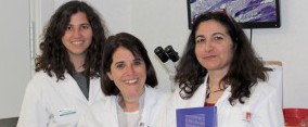
Major grant 2018
"Epitheliotropic lymphoma and interface dermatitis in dogs: identification of discriminatory biomarkers for ambiguous cases"

Grant receivers: Dr. Martina Dettwiler and Prof. Monika Welle, Insitute of Animal Pathology, Vetsuisse Faculty, University of Bern, Switzerland. In the picture is also Nadja Gerber, doctoral student (middle)
Presentation of study: With this study we aim to identify biomarkers reliably distinguishing CETL and IMID in canine skin biopsies with inconclusive histopathological, immunohistochemical and lymphocyte clonality assessment findings, and to establish a protocol for their application in the diagnostic setting.
________________________________________________________________________________________
Training grant 2018
“A comparison of longitudinal and transverse sections in the histological diagnosis of selected canine non-inflammatory alopecic diseases: technique standardization and correlation with dermoscopic findings”


Grant receivers: Elena Borio, Paola Roccabianca, Francesca Abramo, Fabia Scarampella, Francesco Albanese, , Giordana Zanna
Presentation of study: In humans, alopecia can be accurately diagnosed based on clinical presentation and progression of hair loss. Histopathology and dermoscopy are adjunctive aids through provision of further morphological information, and transversely sectioned biopsy specimens provide many advantages in the diagnosis of patients with alopecia. In contrast, only a few studies on the application of dermoscopy and/or transversal histological sections have been documented in veterinary dermatology. According with this background, the aims of the current project are first to evaluate if also in dogs, transverse skin sections can represent a more effective approach to the examination of selected cases of non-inflammatory alopecia when compared to longitudinal skin sections. Secondly, a dermoscopic-histopathological correlation between results will be performed in order to evaluate the role and usefulness of dermoscopy also in these specific canine hair loss disorders.
Minor grant 2018
"A Potential of the Culture Test Flexicult Vet for Canine Pyoderma Management"

Grant receivers: Kaja Winkler, Blaž Cugmas
This award resulted in an article in Antibiotics 2021, 10 (10), 1160; https://doi.org/10.3390/antibiotics10101160"How Accurate Are Veterinary Clinicians Employing Flexicult Vet for Identification and Antimicrobial Susceptibility Testing of Urinary Bacteria?"
Presentation of the study: Antibiotic treatment of pyoderma is often chosen empirically from the list of first-line antibiotics. However, in severe cases of pyoderma (recurrent pyoderma, rods, life threating cases) and when empirical antimicrobial therapy does not resolve the infection as expected, a culture investigation and a susceptibility test for second-line antibiotics are needed. Since the standard antimicrobial susceptibility testing (AST) takes a few days, so-called Point-of-Care tests for AST were introduced. They can be performed at the clinic, reducing turnaround time. One of them is Flexicult Vet, a culture test for diagnosing urinary tract infections (UTI) in dogs and cats. The test includes various antibiotics added into the agar. In the proposed project, we want to test potential of Point-of-Care culture tests for detection and AST of canine pyoderma pathogens. In parallel to AST with Flexicult Vet, all samples will be tested by standard methods. Currently, we are searching for an optimal sampling method.
Practitioner grant :
"Canine flank alopecia"
Grant receivers: Dr. Millie Verschuuren-Tjoeng and Dr Yvette Schlotter


Millie Verschuuren Yvette Schlotter
Primary objective: To determine the preventative effect of subcutaneous slow-release melatonin implants on recurrence of Canine Flank Alopecia (CFA) in dogs with a history of CFA, in comparison to dogs with no treatment.
Secondary objective: To determine the prevalence of CFA in dogs from high risk breeds in the Netherlands.
_________________________________________________________________________________________
Major grant 2017
"Characterization and differentiation of canine hair follicular organoids: an important in vitrotool to study the pathogenesis of non-inflammatory alopecia "

Grant receivers: Dr Dominique Wiener, Dr Monika Welle
Presentation of the study: Non-inflammatory alopecia is a frequent problem in dogs and a frequent reason for consulting a veterinarian. The possible underlying causes are numerous; however, the pathogenesis of most alopecic disorders is still unclear. Hair follicles (HFs) cyclically renew themselves life-long through a pool of HF stem cells (SCs). Accordingly, disturbances in the HF SC pool affect HF morphogenesis, reconstitution or differentiation including the formation of the hair shaft and thus are frequently the cause of alopecic conditions. The underlying causes of an impaired SC function resulting in alopecic disorders remain however to be explored. By expansion of SCs from several adult tissues in vitro 3D “mini-organs”, called organoids, can be cultured. By using culture conditions that manipulate specific signaling pathways detailed analyses of specific pathologies can be explored. Thus besides representing an exquisite tool to eventually generate HFs in vitro, organoids established from HF SCs also bear a great potential to study functional defects in HF morphogenesis, reconstitution and differentiation leading to alopecic diseases and to establish novel therapies. Recently, we achieved to establish organoids derived from canine HFs. These organoids can be kept in culture for up to 16 weeks and can be already used to study the effect of certain signaling pathways. However, the evaluation of several HF specific markers showed that these organoids although derived from HF cells are still lacking a distinct HF signature. Therefore, the aim of this project is to differentiate organoids derived from HFs in different hair cycle stages of healthy dogs into HF organoids with a distinct HF signature. We expect that the results of the proposed project will foster our understanding of normal HF SC biology and in a follow up study these organoids will enable us to unravel the pathogenesis of SC-related alopecic disorders in dogs.
Training grant 2017
"Whole-genome sequencing and comparative genomic analysis of the Staphylococcus isolates from superficial pyoderma forms in dogs"

Grant receivers: Frane Banovic, Walt Lorenz
Presentation of the study: Superficial staphylococcal pyoderma is a common disease in the dog characterized by three clinically and histopathologically distinctive forms: superficial bacterial folliculitis (SBF), bullous impetigo (BI) and exfoliative pyoderma associated-epidermal collarettes (EC). These conditions are frequently recurrent and difficult to treat due to the worldwide emergence of methicillin and multidrug-resistant strains; Staphylococcus (S.) pseudintermedius is the major pathogen. The knowledge of genetic elements responsible for the virulence and the metabolic pathways utilized by pyoderma-associated staphylococci is limited. We propose to evaluate the molecular pathogenesis of the main staphylococcal pyoderma phenotypes; we wish to determine the whole-genome sequences and perform a detailed genomic comparison analysis between pyoderma-associated S. pseudintermedius strains collected from each of the different superficial pyoderma forms (SBF, BI, EC) to identify the genes likely associated with lesion development. These bacterial isolates represent a spectrum of antibiotics resistance from sensitive to all antibiotics to multidrug-resistant, including methicillin. To determine the staphylococcal whole genomes, a total of 12 previously acquired, stored S. pseudintermedius isolates (four different isolates from SBF, BI and EC lesions) will be sequenced at our institutional Genomics Facility using an Illumina MiSeq 600 cycle run. Whole genomes will be annotated to allow functional gene membership identification (e.g genes encoding toxins or adhesion) for all strains. To assess differences in virulence factors and metabolic pathways, whole genome circular comparative maps will be generated and differentially expressed subsystems will be clustered to reveal strain-specific subsystems. Finally, we will reconstruct strain-specific metabolic pathways for each strain. We hypothesize that the molecular characteristics of S. pseudintermedius superficial pyoderma-associated strains are different between pyoderma phenotypes, particularly the virulence factors associated with colonization fitness, disease pathogenesis and regulation of virulence factor expression.
Practitioner grant 2016
"Role of elastography in the evaluation of canine skin: principles and clinical considerations”
Grant receivers: Eric Zini, Giordana Zanna, Giulia Brizzi, Edoardo Auriemma and Simona Morabito
Presentation of the study: Elastosonography is a painless, invasiveness and promising imaging method that allows the assessment of tissue elasticity. It is based on the principle that softer, normal tissue displaces more easily than harder, malignant tissue when an oscillatory pressure is manually applied by an ultrasound transducer. The variation in deformity is seen as changes in ultrasound signals which are represented on the video screen by a color map called elastogram. The increased tissue hardness appears in ascending order as red, yellow, green and blue. In human medicine, elastosonography is currently used in several cutaneous and internal organ disorders as an intriguing alternative to the traditional diagnostic tools because it provides useful information about the stiffness and the anatomic features of a lesion. In dermatology, it has been used to evaluate cutaneous neoplasms as melanoma and carcinoma or diffuse cutaneous systemic sclerosis and lymphedema. In veterinary, few reports are reported on the application of elastosonography on several tissues as liver, splenic and renal parenchyma or in mammary tumors. However, no studies have been performed about the use of elastosonography in veterinary dermatology. The aim of this project is to correlate elastosonographic with histopathological findings of nodular lesions in dogs, in order to identify elastosonographic criteria that may provide further information to the clinician in the morphological screening of these lesions.
Major grant 2015
"Evaluation of the cutaneous immunological milieu and leptin expression in dogs naturally affected by Leishmania infantum/chagasi before and after meglumine antimoniate treatment"

Grant receivers: Dr Antonio Di Loria, Dr Domenico Santoro
Presentation of the study: Leishmaniasis is a wide spread infectious disease endemic in the Mediterranean area for both dogs and people. Although not completely elucidated, an alteration of the cutaneous immune response has been demonstrated in affected dogs. Multiple immunological cell types have been involved in the immunological response in affected animals. It is well known that a cell-mediated response (T helper [Th] 1 type) is the cornerstone of dogs’ resistance against the parasite; however no studies have been reported on the immunological status (innate and adaptive immunity) of naturally affected dogs before and after treatment with meglumine antimoniate. In people with cutaneous leishmaniasis treated with meglumine antimoniate, it has been shown that an enhancement of the phagocytosis and TNF-α production by peripheral monocytes is present compared with monocytes not exposed to antimoniate. No information is currently available on the involvement of innate immunity in canine leishmaniasis; the only exception is the proven in vitro efficacy of antimicrobial peptides (AMPs) against Leishmania parasite. In addition, very few biological markers, to rapidly identify the immunological response, are available in veterinary medicine. In people, biological molecules able to orchestrate the Th1 and Th2 immune response and their potentials for therapies against infectious agents have been investigated in the past few years. One of such markers is leptin, a hormone secreted primarily by adipocytes and immune cells. Very recently it has been shown that an increase in leptin mRNA expression is present in peripheral blood mononuclear cells in canine leishmaniasis compared with healthy dogs. This study supported the idea of leptin as possible marker of severity and prognosis in affected dog